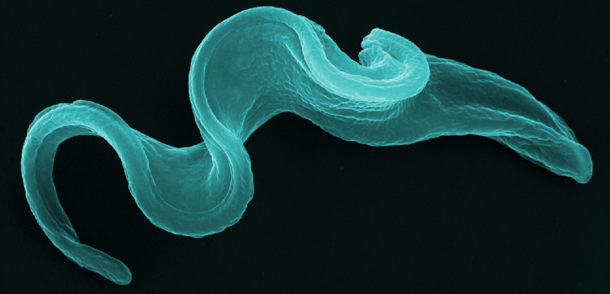
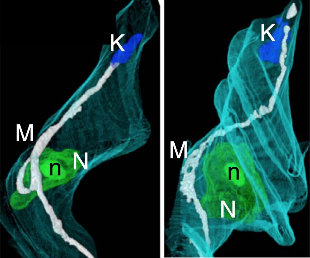
FIB-SEM tomography of wild-type Trypanosoma brucei. In this type of tomography, 5-nm layers of the surface (∼2 µm2) of a plastic block containing heavy-metal-stained trypanosomes, are abraded by an ion beam (FIB) and the exposed surface imaged by Scanning Electron Microscopy (SEM). Thousands of images are then compiled and reconstructed into a 3D volume. M, mitochondrion; K, kinetoplast (mitochondrial DNA); N, nucleus; n, nucleolus; turquoise, parasite’s surface. VIDEO
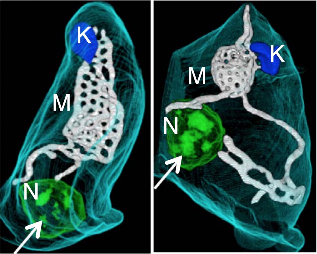
FIB-SEM tomography of APOL1-treated wild-type Trypanosoma brucei. M, mitochondrion; K, kinetoplast; N, nucleus; turquoise, parasite’s surface. Notice the mitochondrial fenestrations induced by the APOL1 treatment (compare with WT image above). VIDEO
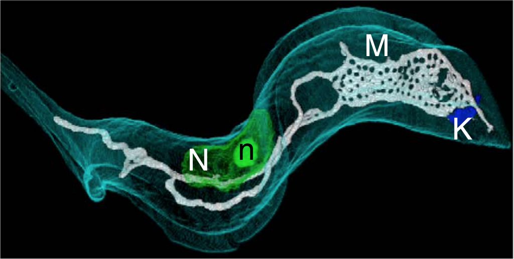
FIB-SEM tomography of a RNAi cell line of Trypanosoma brucei ablated for the expression of TbMFL1. M, mitochondrion; K, kinetoplast; N, nucleus. Turquoise, parasite’s surface. Notice the mitochondrial fenestrations induced by APOL1 treatment (compare with WTimage). VIDEO.
Credit: FIB-SEM 3D reconstruction and videos, ©Frédéric Fontaine.
Original reference:
Coupling of lysosomal and mitochondrial membrane permeabilization in trypanolysis by APOL1.Vanwalleghem G, Fontaine F, Lecordier L, Tebabi P, Klewe K, Nolan DP, Yamaryo-Botté Y, Botté C, Kremer A, Burkard GS, Rassow J, Roditi I, Pérez-Morga D, Pays E.Nat Commun. 2015 Aug 26;6:8078. doi: 10.1038/ncomms9078.